
Parts of the Eye
Human eyes are extremely complex. They consist of various components that work with the brain to form vision.
How the Eye Works
Different parts of the eye all work together to make up vision. The major parts include the lens, retina, and optic nerve. Together, those parts transform the light that comes into your eye to images.
Eye Parts and Functions
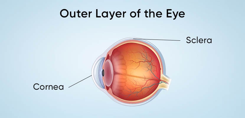
The outer layer of the eye is called the fibrous tunic and consists of the following parts:
- Sclera: This is the strong, white-colored layer of tissue that covers almost the entire surface of the eyeball. The extraocular muscles are connected to the sclera and they control the eye’s movement.
- Cornea: The clear, outermost layer of the eye is the cornea. Its dome shape bends the light that enters the eye.
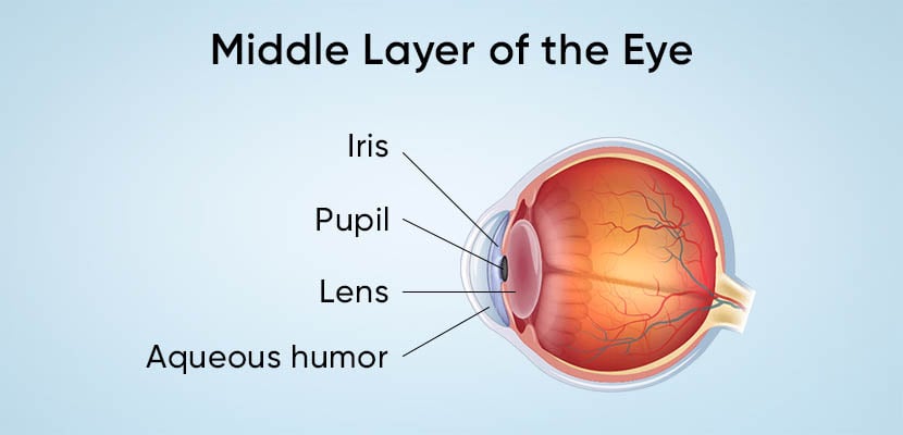
The middle layer of the eye is called the vascular tunic, also referred to as the uvea, and consists of the following parts:
- Iris: The iris is the colored part of the eye containing sphincter and dilator muscles. These control the size of the pupil and the amount of light entering the eye.
- Pupil: The black center of the eye is the pupil. It’s controlled by the iris and is where light enters the eye. Dim light makes it expand and bright light causes it to shrink.
- Lens: The lens focuses light onto the retina and is located behind the pupil. As people age, the lens can become cloudy, which is known as cataracts.
- Aqueous humor: Clear fluid between the lens and the cornea is known as aqueous humor. This fluid helps maintain the pressure inside the eye. When the eye is unable to regulate intraocular pressure (IOP), the high pressure can lead to damage to the optic nerve resulting in glaucoma.
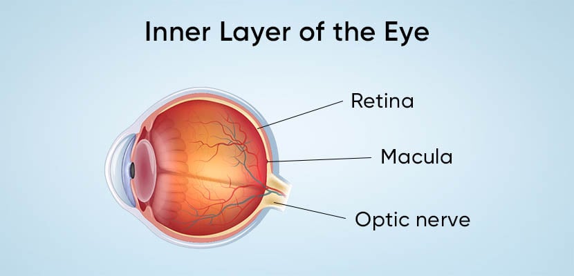
The inner layer of the eye is called the nervous tunic, also referred to as the retina, and consists of the following parts:
- Retina: The layer of tissue in the back of the eye is the retina. The retina contains cells called photoreceptors, known as rods and cones, which transform light focused by the lens into electrical signals.
- Macula: The macula is part of the retina that is responsible for the sharpest portion of our vision. As we age, our eyes begin to weaken, which can result in damage to the macula. This is known as age-related macular degeneration.
- Optic Nerve: The optic nerve sends the electrical signals from the retina to the brain, allowing it to form the images that you see.
Since there are so many parts that work together to make up your vision, there will also be a few common eye problems that can occur. That’s why it is important to schedule annual eye exams and talk to your eye doctor if you notice any changes in your vision.